Defibrillation, DC Politics and Publicity in Heart Transplantation

John A. Macoviak, MD, is a Google Scholar and retired heart transplant surgeon based in California. John Macoviak, MD, now focuses on his work as an author of non-fiction and fiction. In this article John A. Macoviak writes about something good from the medical device world after the approval in 1995 by the FDA of publicly available defibrillators, methods and apparatus used for defibrillating a heart, a great lifesaving advance. Internal surgically implanted cardiac defibrillators and more depth on the often-brutal controversies in heart transplantation which have been and will be further addressed in other articles.

Dr. John A. Macoviak writes on range of medical topics like heart transplantation and Washington DC POLITICS which created powerful media attention at the Heart Transplant Consortium of 7 Hospitals. His mentor Dr. Norman E Shumway was a powerful master of publicity in Palo Alto and loomed large in the small world of heart surgeons globally enshrined in history. Shumway put Dr. Macoviak in the media spotlight when a boy from Poland was flown in for complex congenital heart surgery. Shumway assigned Macoviak to address some of his famous successful outcomes and referrals including 30 heart transplant recipents for all walks of life, and others who sought Shumway’s considred opinion including Edward Teller, Jay Pritzker Sr., Milton Friedman.

The young new surgeon was a never before politician, not of billionaire family heritage unlike his peer co-chief resident Bill Frist. By birth Dr. Macoviak grew up in a coal patch town of less than 500 people in Harwood, Pennsylvania. He had strong genes of resiliency for serving others at all costs like his humble devout parents, his father a WWII Master Sergeant who made sure his 65 men came home honorably, safely and heroically. He never wanted to discuss that. His son was always striving to a fault to make an industrious earned positive difference for family, mentors, friends and patients. John A. Macoviak as a newly trained heart transplant surgeon was retained by the most affluent DC paradigms, leaders and systems, pressured and obliged to follow educate-the-public by requests of the media and Consortium caught up in the competitive town-gown syndrome dating centuries past.

By DC politics and 7 hospitals all wanting recognition, Georgetown, Dr. Macoviak’s college Cum Laude and medical school Magna Cum Laude alma mater was precluded from being the sole heart transplant center. Instead the consensus newly default created in 1988 the Seven Hospital Washington Regional Heart Transplant Consortium. In the consortium Macoviak performed the first thirty heart transplants, one compassionate experimental heart pancreas transplant on a diabetic nuclear physicist having normal kidneys with Hans Sollinger MD the world’s leading pancreas transplant surgeon at the University of Wisconsin. All plans of action were approved by the full Ethics Committee and co-surgeons Department Chairs Paul Corso MD (Cardiac Surgery) and Jimmy Light MD (Renal Transplant Surgery).

Dr. Macoviak also performed the first LVADs in the US after Texas Heart at Medstar. Also he performed one heart transplant at GW with Ben Aaron, and one heart transplant at Georgetown with Nevin Katz MD. John A. Macoviak writes often about experiences on faculty at Harvard, Stanford, Mayo, Georgetown and Penn and their world-renowned Professors of Cardiology and Cardiac Surgery. He was fortunately blessed by great mentors who without exception always fully supported Macoviak without reservation publicly and privately in the face of archetypically outwardly-humble but quietly-operating clandestine competitor contrarians, whom his daughters said “they had to – they all wanted to be you” and “because it was a very challenging exhausting against all odds nonetheless successful important legacy, built standing on the shoulders of great mentors.”

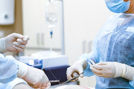
Turning to less controversial life-saving technology advances “Defibrillation”, is alternatively referred to as electrical cardioversion, a medical procedure that uses an electrical current to modify a person’s heartbeat, with the goal of returning the heart to a normal, healthy rhythm. Various conditions and medical events can result in potentially fatal arrhythmias in the lower chambers of the heart, which must be remedied through defibrillation.

While defibrillation is an effective strategy for addressing abnormal heart rhythms, medical professionals will have more success with the technique if they apply it as soon as the arrhythmia develops. Defibrillation technology can manage a variety of abnormal heartbeats, including ventricular tachycardia (defined by a quick heartbeat with no pulse) and ventricular fibrillation (a quivering heart muscle resulting from cardiac arrest).

While defibrillators are often used during medical emergencies, individuals living with potentially fatal heart conditions must carry personal defibrillators with them at all times. These devices range in form from a vest-like, wearable defibrillator to implantable cardioverter defibrillator devices, which are comparable to a watch.

While events requiring defibrillation may be frightening, defibrillation is fairly common in the United States, Every year, about 200,000 hospital patients and 184,000 individuals outside of hospital settings experience cardiac events that require defibrillation.

How Do Surgeons Actually Practice Surgery?

john macoviak

Surgeons are often viewed as an elite tier of doctors renowned for their steady hands, surly dispositions, and eyes for detail. Unlike other fields of medicine, though, surgeons cannot practice their craft by passively studying books or speaking with patients. They must actively hone their skills into an art form. In the following article, John A. Macoviak answers the question: how exactly do surgeons practice surgery? “Surgery is not hard to do, its hard to get to do” said one once famous then infamous surgeon who brutalized trainees. “If the resident isn’t in the inner circle they are often simply worked to exhaustion assisting, taking night calls, rarely being allowed to do any of the procedures”. The theory is the Program Directors only count on 1 in 10 rising to the top preferable from wealthy stock. The others are needed to run the ship but are cut off from honing skills especially if they deviate accidentally or by their genetic back grounds in any way from the normal expected servitude. That is referred to as pyramid programs whether formal or clandestine.”  

In this post, we’ll explore how medical students slowly train to become surgeons and discuss how they practice cutting into bodies without harming patients. We’ll also touch on the role of technology in modern surgery and how it has allowed surgeons to become even more skilled at their jobs.

How Students Transform into Surgeons

The first step in becoming a surgeon is, of course, to attend medical school. During their time in medical school, students will take classes on human anatomy and physiology. They will also learn about the different surgical procedures they’ll be expected to perform but the majority of their time will be spent in the classroom, not in an operating theater.

After medical school, surgeons-in-training will complete a residency program. This is where they will finally get some hands-on experience in the operating room. During their residency, they will work closely with experienced surgeons and learn how to perform different types of surgeries. They will also learn how to handle different types of medical emergencies.

After completing their residency, surgeons will take a series of exams to become certified by the American Board of Surgery. Once they are certified, they can finally start practicing surgery on their own.

Of course, becoming a surgeon is not just about acquiring the necessary skills. It is also about having the right mindset. Surgeons need to be able to think quickly and make decisions under pressure. They also need to have a high tolerance for stress. So, how can surgeons practice and hone their skills without performing a real surgery?

Cadavers

While training in medical school, most students learn to perform surgeries by operating on a cadaver. This gives them hands-on experience with a real human body, allowing them to see real organs and tissues and learn to implement different surgical procedures. Yet, as technology has advanced, most med schools have acknowledged the limitations of using cadavers:

  • Cadavers are often expensive due to a limited supply.
  • Cadaver-based skills training doesn’t help surgeons learn to identify or repair bleeds or damage to blood vessels.
  • Cadavers do not show signs of life, so students cannot learn to assess a patient’s condition during surgery.

For these reasons, most students learn to perform surgeries using a blend of methods, not just cadavers.

john macoviak

Simulated Surgeries

As augmented and virtual reality tech has become more advanced, the medical field has quickly adapted to incorporate these features into surgical training programs. Rather than relying entirely on cadaver surgeries, many medical schools are now turning to virtual reality (VR) simulations as a way to provide students with more realistic training experiences.

VR simulations can be used to create realistic 3D models of human anatomy, which can be used to teach students about specific organs and tissues. In addition, VR simulations can be used to create realistic surgical scenarios, allowing students to practice different procedures on virtual patients.

There are a number of different VR platforms that are being developed for use in medical education, but the two most promising platforms are Osso VR and the Oculus Right. Oculus Rift is a VR headset that can be paired with software developed through a joint effort between Johnson & Johnson and Osso VR. It includes a number of different medical simulations, including heart surgeries and childbirth simulators.

Baptism by Fire

Once surgeons leave medical school and finish their residencies, they’re unlikely to ever practice on a cadaver or in a VR simulation again. Instead, they’ll gain all of their experience by operating on real patients. This is why it’s essential that surgeons gain as much simulated experience as possible before they graduate.

If a surgeon makes a mistake during a real surgery, it could have dire consequences for the patient. At the very least, they could put themselves at risk of a malpractice suit. Nonetheless, they call medicine a practice for a reason. Every time a surgeon picks up a scalpel, they further hone their well-tempered skills into an art form.

Final Thoughts

Surgeons spend many years developing the skills and knowledge needed to operate on real patients. By practicing on donated cadavers and in VR simulations, surgeons progressively learn to perform complex surgeries that could kill their patients. After graduating and finishing a residency, surgeons then continue to practice their skills every time they enter the operating room.

Moderate Alcohol Consumption Can Increase the Risk of Hypertension

A retired heart transplant surgeon in California, John A. Macoviak, MD, focuses his time on being a medical author and blogger. Having written more than 50 peer-reviewed articles, along with performing many ground-breaking surgeries, John Macoviak MD MBA maintains a professional interest in both hypertension and diabetes.

According to research from September 2020, adults who have type 2 diabetes (T2D) are more likely to develop hypertension with moderate alcohol consumption. The study, which was published in the Journal of the American Heart Association, looked at data from 10,200 adults participating in the ACCORD trial. This trial previously compared methods of reducing the risk of cardiovascular disease in adults who had T2D.

For the current research, adults were split into three categories: those who were light drinkers, moderate drinkers, and heavy drinkers. Light drinkers only had one to seven drinks per week, where each drink was 12 ounces of beer, 1.5 ounces of liquor, or six ounces of wine. Meanwhile, heavy drinkers had upwards of 15 drinks per week, while moderate drinkers had between eight and 14 drinks.

Based on the study’s analysis, heavy drinkers had almost twice the risk of elevated blood pressure (BP) readings. They were also three times more likely to develop stage II hypertension and 2.5 times more at risk of stage I hypertension. For moderate drinkers, the risk dropped, but it was still significantly above their peers who drank less.

Percutaneous Septal Sinus Shortening for Functional MR

A retired heart transplant surgeon and researcher, John Macoviak MD MBA currently writes on a range of medical subjects, with special focus on the treatment of heart conditions. Among his previous research publications is the paper titled “Percutaneous septal sinus shortening: a novel procedure for the treatment of functional mitral regurgitation.”

The most common valve disease, functional mitral regurgitation, also known as secondary mitral regurgitation, is characterized by motion abnormalities in the wall of the left lower chamber of the heart (left ventricle), compromising closure of the mitral valve (the valve between the left upper and lower chambers of the heart). Mitral regurgitation causes the left ventricle of the heart to become susceptible to further damage, since the volume of blood it holds is higher than normal due to reverse flow of blood past the mitral valve. This condition can eventually cause death if not treated. Functional MR happens when other diseases of the left ventricle have resulted in severe left ventricular dysfunction. Often, it is inadvertently discovered during a patient’s examination for heart failure. Among other symptoms, patients with functional mitral regurgitation usually experience shortness of breath.

In the publication, John Macoviak and other researchers describe the percutaneous septal sinus shortening system for treating functional MR. This novel technique was applied to a sheep model, and the result showed significant success. This technique reduced the annular diameter of the mitral valve from center to the left in sheep with severe functional MR to a diameter optimal for reducing mitral regurgitation. While the study employed a standard catheter technique, the researchers noted that the technique could further be enhanced by using fluoroscopy.

Cortisol Levels Linked to Blood Sugar in People with Diabetes

John Macoviak MD MBA, a graduate of Georgetown University, is a retired Washington, DC, heart transplant surgeon. Now living in California, John Macoviak MD MBA continues to write about medical conditions, most notably chronic conditions like diabetes.

The results of a new study by researchers at the Ohio State University College of Medicine and the Ohio State University Wexner Medical Center has revealed a link between blood sugar and cortisol levels in people living with type 2 diabetes.

Published in Psychoneuroendocrinology, the July 2020 study builds on previous research that showed both stress and depression lead to more stable cortisol levels throughout the day. These sustained levels are not seen in healthy people, for whom cortisol naturally is high at the beginning of the day before dropping in the evening.

Since cortisol is linked to a decrease in insulin and an increase in blood sugar, having sustained levels makes diabetes much more difficult to control. This is why it’s essential that people with type 2 diabetes find ways to minimize their cortisol levels so that they do not experience blood sugar spikes.

To find this link, researchers examined data from more than 2,000 participants between the ages of 45 and 84. These participants were followed for a period of six years, during which time researchers noticed that people with diabetes had steadier cortisol levels and higher glucose levels than their peers without diabetes.

How Depression Increases the Risk of Heart Disease

A retired heart transplant surgeon and dedicated medical researcher, John Macoviak MD MBA has been a respected member of the medical community for more than four decades. Passionate about sharing his medical expertise with the public, John Macoviak MD MBA actively writes articles about different medical conditions. This includes an online article he wrote discussing heart disease in women, and common risk factors such as depression.

When people are depressed, they often experience low-grade inflammation, which contributes to the clogging of arteries. Beyond that, people who are depressed experience a higher production rate of stress hormones. Stress hormones contribute to artery blockages by activating platelets in the blood and making them more likely to clot. Such hormones also limit how much the arteries and heart respond to requests for increased blood flow in the body. Both factors increase the risk of heart disease in people with depression.

Depression increases the risk of heart disease in other ways as well. People who are depressed commonly experience lower energy levels or a lack of motivation. As a result, they struggle with eating healthy foods and maintaining a regular exercise routine. Such lifestyle choices are essential for reducing various cardiovascular and other health issues.

This change in motivation and energy additionally leads to disruptions in people’s medication behavior. They may take medicines incorrectly, or skip taking their medications altogether. Further, people who are depressed have a higher likelihood of self-medicating with alcohol or smoking.

American Institute of Biological Sciences Advocates for Science

A Magna Cum Laude graduate of Georgetown University School of Medicine, John Macoviak MD MBA taught medicine at esteemed universities, including the the Hospital of the University of Pennsylvania, Stanford Hospital, Georgetown University Hospital, Harvard School of Medicine and Mayo Clinic. John Macoviak MD MBA has spent time working on significant research projects, including serving on NASA’s space medicine peer review panel through the American Institute of Biological Sciences, a scientific association aimed at growing knowledge of life sciences.

Established in 1947, the American Institute of Biological Sciences originated as a part of the National Academy of Sciences, but would go on to become an independent organization in the 1950s. The membership consists of more than 250,000 biologists and scientific professionals.

The American Institute of Biological Sciences pursues its goals of broadening life science education through events and activities meant to influence public policy. The organization leverages its location in Washington, D.C. to advocate for the scientific community with governmental representatives.

How Cytomegalovirus Can Affect Adults

A retired heart transplant surgeon in California, John Macoviak MD MBA is an esteemed member of the medical community. Since performing his first heart transplant in Washington, DC, he has performed several groundbreaking surgeries and written upwards of 50 medical and surgical articles. John Macoviak’s publications have covered such medical conditions as cytomegalovirus (CMV) following heart-lung transplantation.

Related to the herpes virus, CMV is an extremely common infection. In the United States, anywhere from 50 percent to 85 percent of adults have the virus. Meanwhile, nearly all adults have CMV in developing countries. Part of why the virus is transmitted so easily is due to the fact that it does not cause serious symptoms in those with healthy immune systems. In fact, many people do not experience symptoms of the virus at all. If they do, they have a low-grade fever that lasts for a few days or weeks, decreased appetite, and fatigue.

However, this isn’t the case for people with weakened immune systems. This includes people who have immune-suppressing diseases, like AIDs, those who are receiving chemotherapy, and those who have had transplants of bone marrow or organs. In these individuals, CMV is linked to retinitis, an eye condition that leads to blindness, or issues with the gastrointestinal tract. Parts of the nervous system and the brain can also be affected by CMV when the immune system is weakened, thus leading to leg weakness and confusion.

Hemiarch Aortic Perfusion Reduces Brain Emboli

Doctor consulting with patient presenting x-ray film results on digital tablet tablet sitting Premium Photo

A graduate with distinction from Georgetown University School of Medicine, John Macoviak MD MBA is a retired cardiothoracic surgeon. John Macoviak MD MBA has researched a wide variety of heart-related conditions, and is a co-author of several medical papers, including “Hemiarch aortic perfusion decreases brain emboli in beating heart surgery.”

An abbreviation of beating heart surgery, BHS refers to any surgery that is performed on the heart while it is still beating which avoids cross clamping the aorta as is usually done with the heart at rest. . One of the main concerns of doctors performing heart surgery is the maintenance of blood circulation (perfusion) during the procedure using the heart lung bypass machine. Aortic cross clamping to stop the heart can result in the formation of cholesterol emboli. Emboli are obstructions in blood vessels that can be also caused by air bubbles, blood clots, or other matter in the blood..

Research was conducted on 13 pigs to study the effect of directing cold blood perfusion in the aorta to the brain to prevent embolism damage to the brain. The aorta is the largest artery in the body, emanating from the upper left chamber of the heart to form an arch in the chest, with branches that convey blood to the brain and other parts of the upper body as well as lower parts of the body. .

During the research, tiny colored microspheres (15 micrometers in diameter) were injected into the base of the aorta and left upper heart chamber of some test subjects. A group of the test subjects had dual blood lumen catheters inserted into the base of the aorta to help channel blood from the upper lumen into the aorta with cold blood, and the lower lumen with warm blood to the rest of the body. While another group had no catheter (to serve as control). At the end of the research, the group with the catheter perfusing cold blood to the brain had fewer emboli going to the brain as compared to the control group.

Electrical Conditioning of Skeletal Muscle for Myocardium Replacement

A graduate of the Georgetown University School of Medicine, John Macoviak MD MBA is a former associate professor of cardiothoracic surgery at Harvard Medical School. John Macoviak MD MBA co-authored articles in several medical publications, including “Electrical conditioning of in situ skeletal muscle for replacement of myocardium.”

Also referred to as the cardiac muscle, the myocardium constitutes most of the heart and is known for the role it plays in the contraction of the heart. Due to the important roles performed by these cells, death of cardiac muscles in the heart can result in complications. To address these cases, surgical intervention may be necessary. Some approaches rely solely on the replacement of dead cardiac muscle cells.

Research conducted in-vivo investigated the efficacy of replacing dead cardiac muscle cells with electrically stimulated skeletal muscle cells from the right diaphragm of test subjects. Skeletal muscles are different from cardiac muscles and are responsible for voluntary movements. During the study, electrical frequencies of 2Hz and 10Hz were used for stimulation over a period of 135 days. The researchers discovered that almost all fast-twitch and easily fatigued muscle cells (known as type I) transformed into slow-twitch and fatigue-resistant muscle cells (type II) with a significant increase in the thickness of stimulated muscles.

Muscle cells that were electrically stimulated at 10Hz contracted vigorously and showed no symptoms of fatigue throughout the study. The conclusion suggested that skeletal muscles of the diaphragm may be a more suitable autograft (grafts gotten from recipient) for replacement of defective or dead cardiac muscle cells.

Design a site like this with WordPress.com
Get started